
Real work. Real clinic.
Cases treated at our Beit Shemesh clinic, and the room you'll actually sit in. No stock photography.
Treatment we've carried out
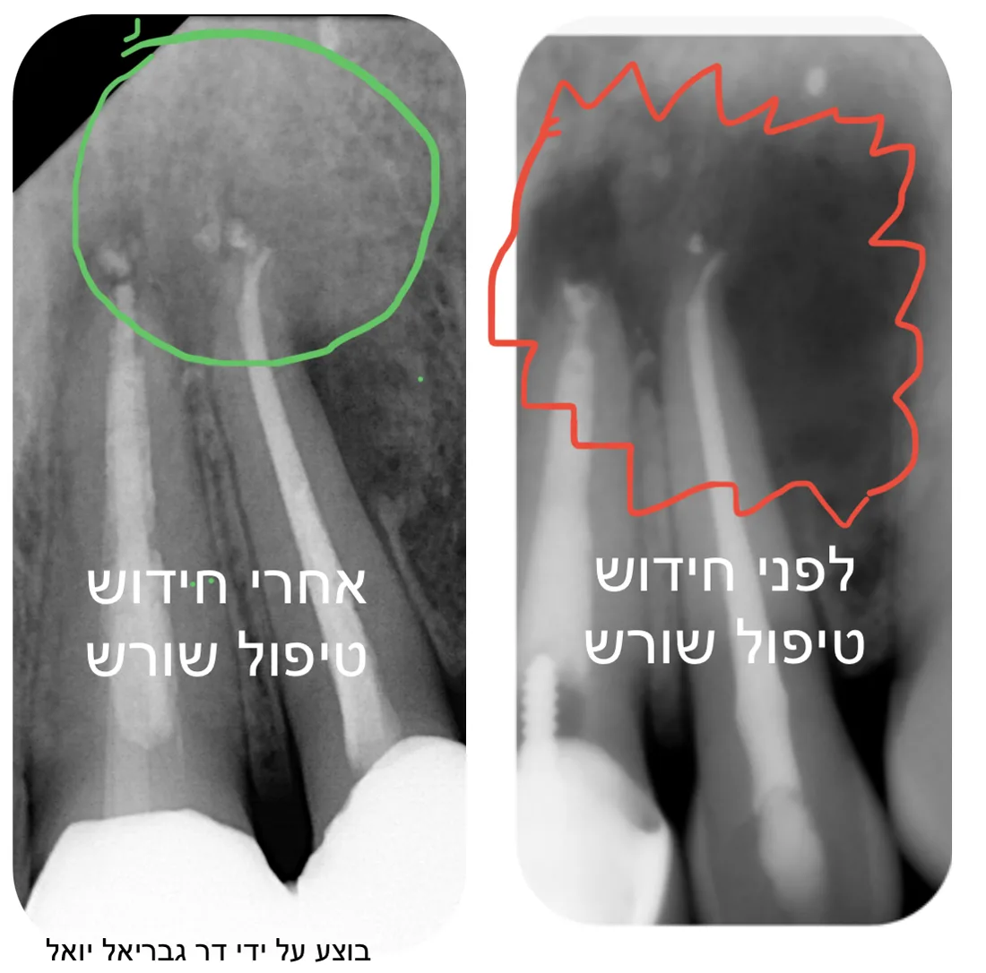
Root canal retreatment
Before and after retreatment of a root canal that had been treated previously. Performed by Dr. Gabriel Joel.
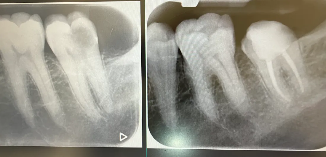
Before and after, side by side
The same back tooth before and after root canal treatment — the filled canals are the bright lines running down the roots.
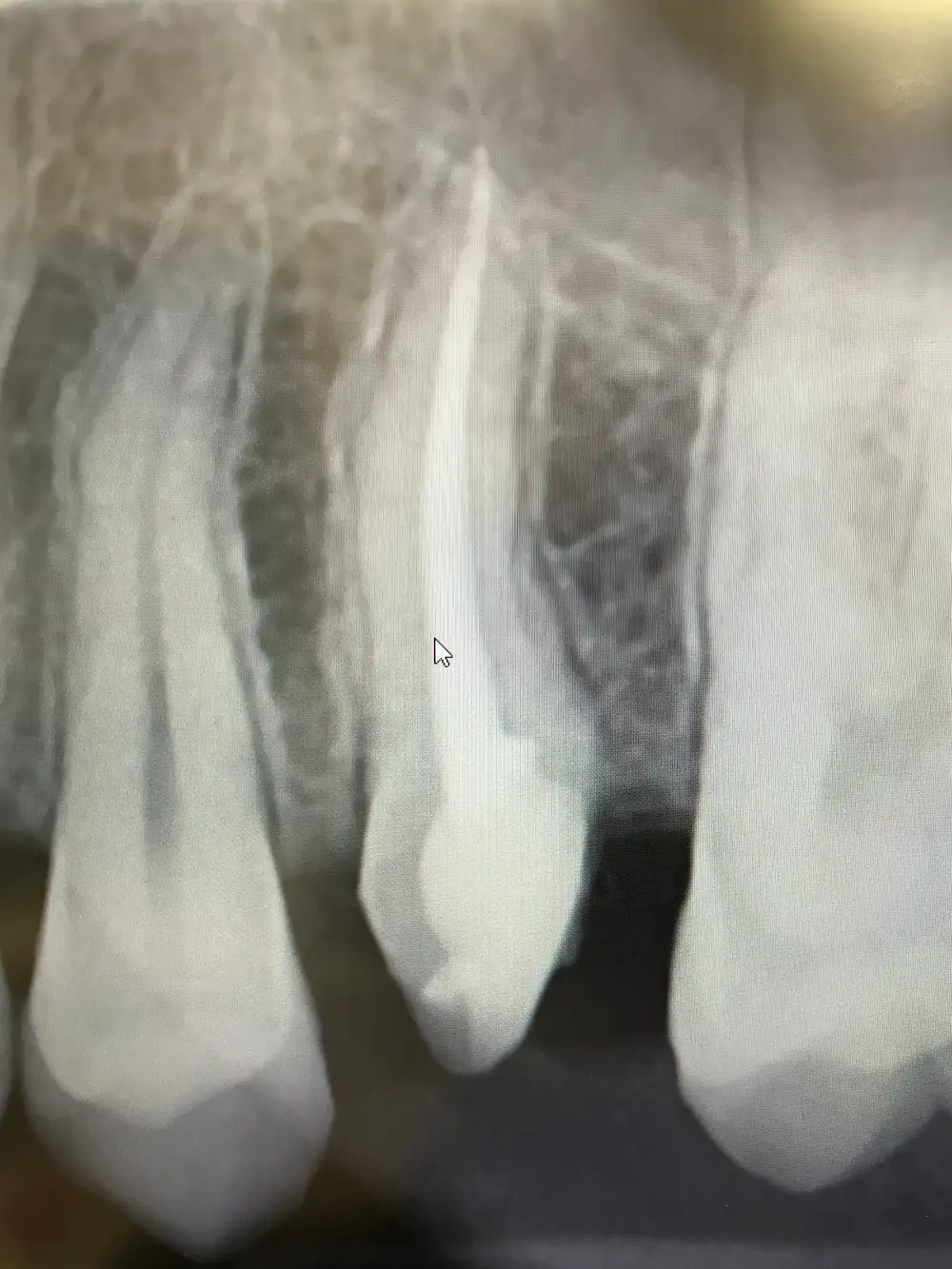
Filled to the root tip
A completed root canal on a single tooth. The canal is sealed the full length of the root.
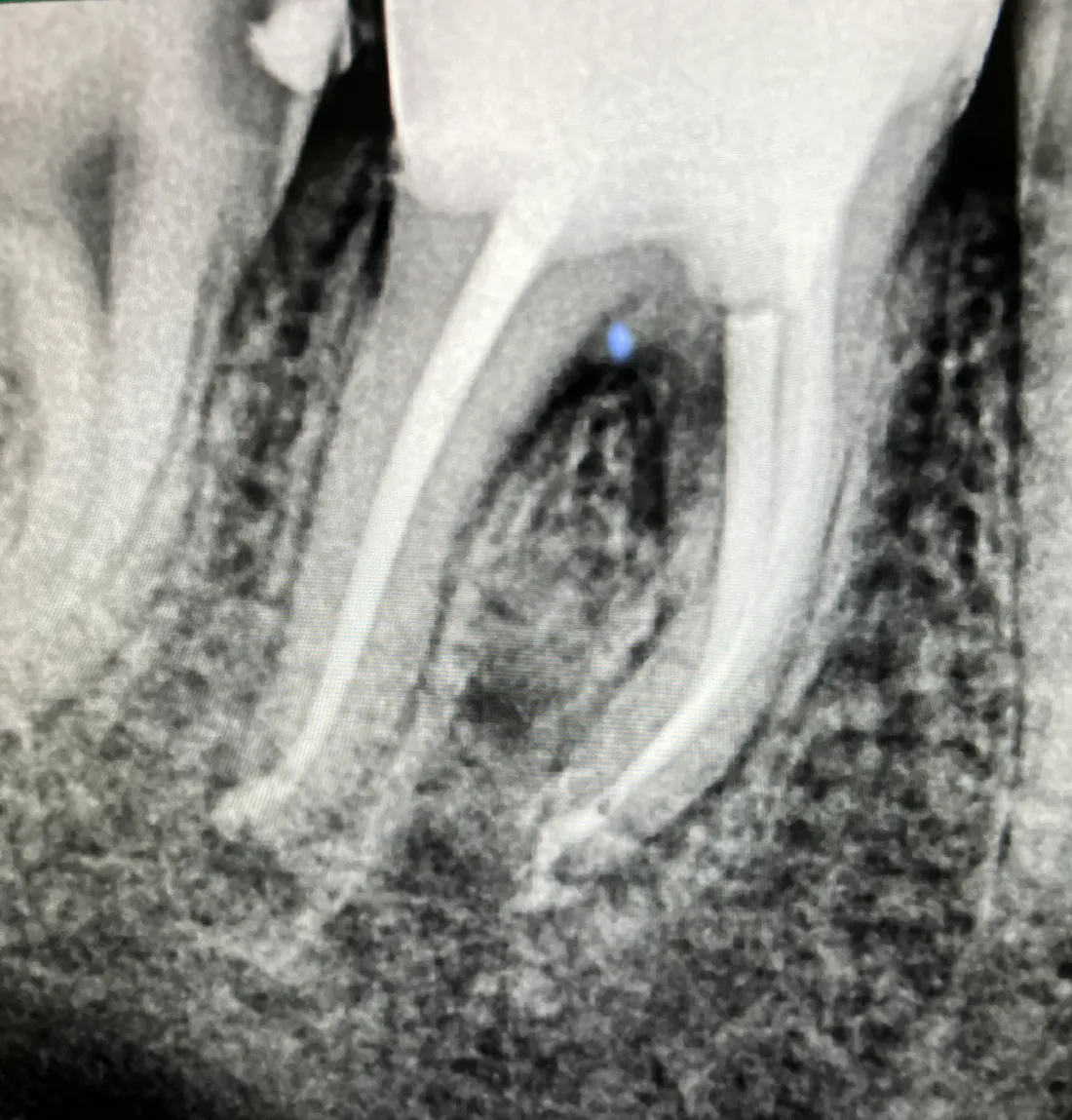
Curved canals in a molar
Back teeth often have narrow, curved canals. Both were cleaned and filled along their full curve.
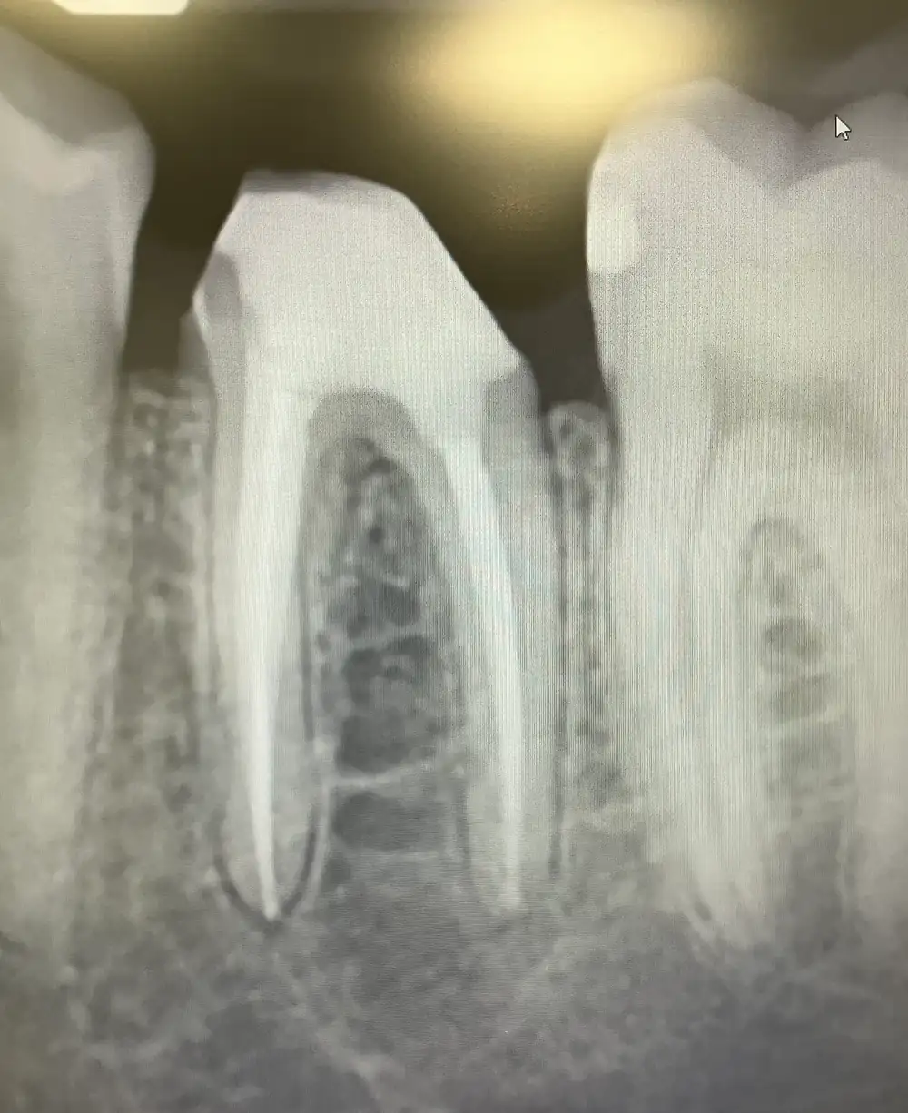
Lower back tooth
Root canal treatment completed on a lower back tooth, keeping the natural tooth in place.
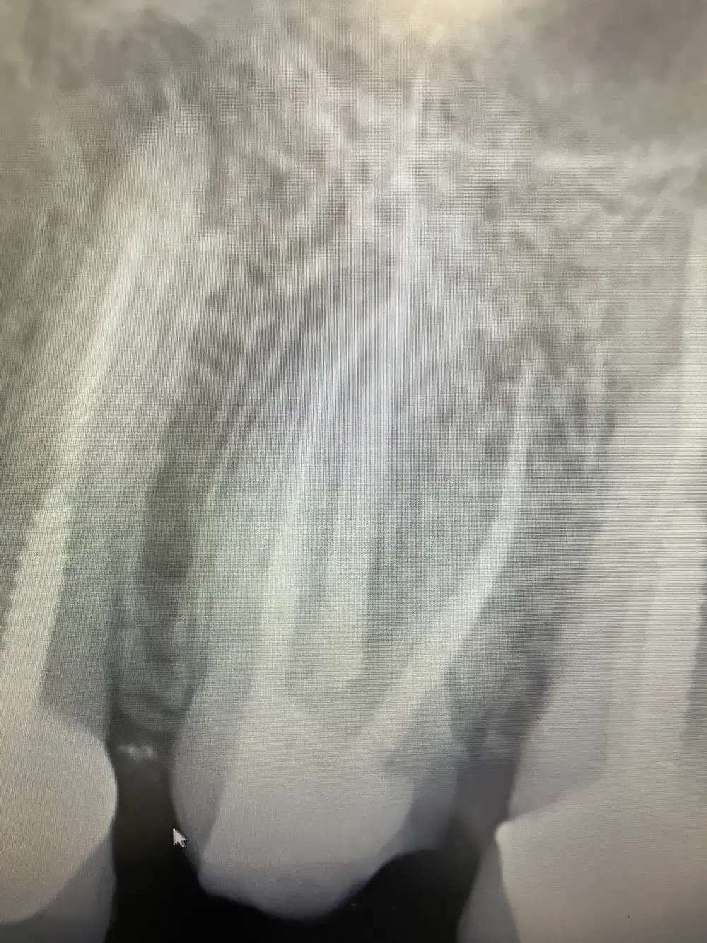
Upper front teeth
Root canal treatment on upper teeth — the filled canals run the length of each root.
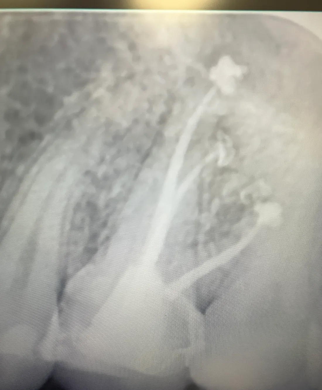
Upper molar
An upper molar treated and sealed. Molars carry the most chewing force, so saving them matters.
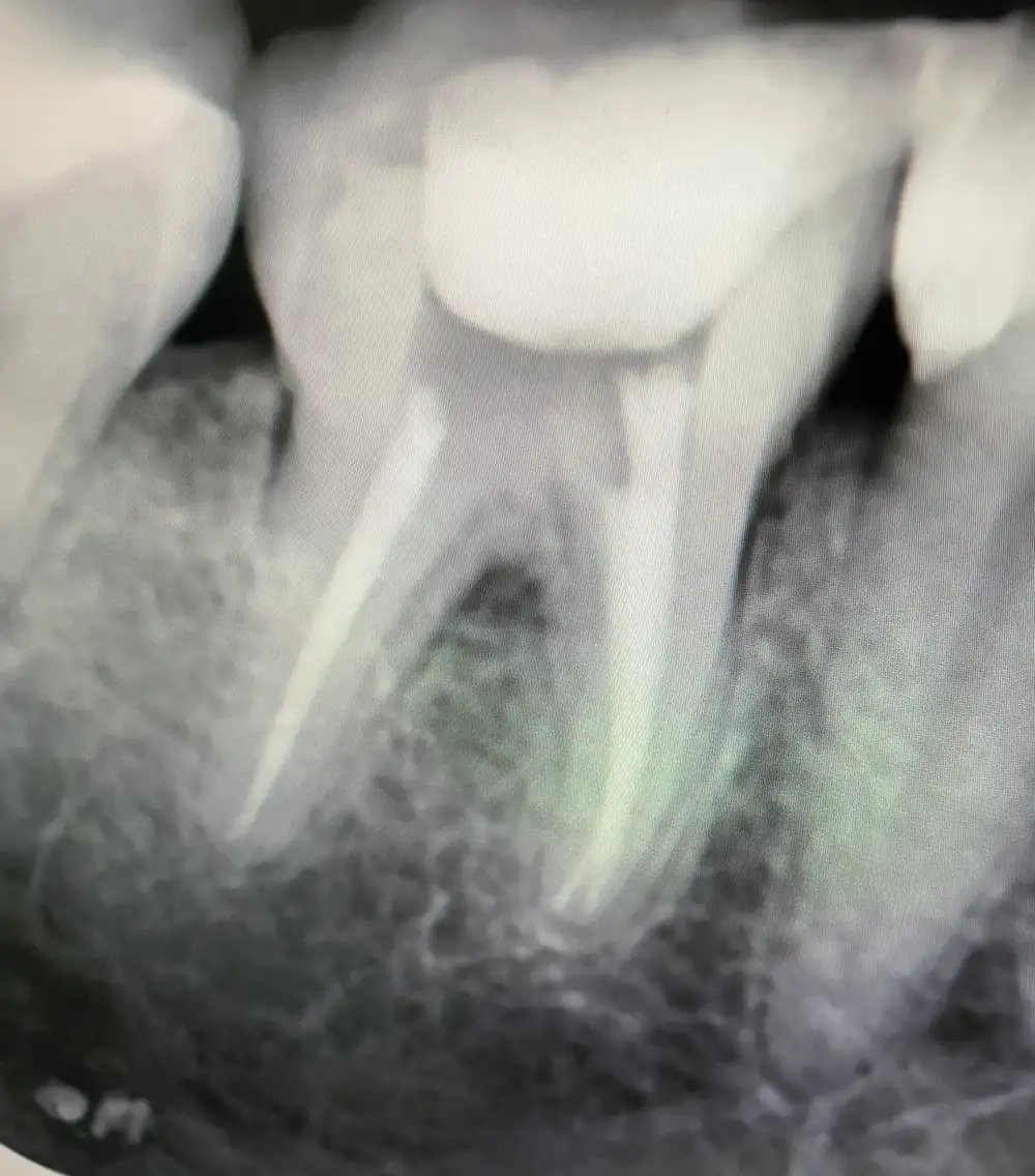
Both canals sealed
Both canals of a lower molar cleaned and sealed in the clinic.
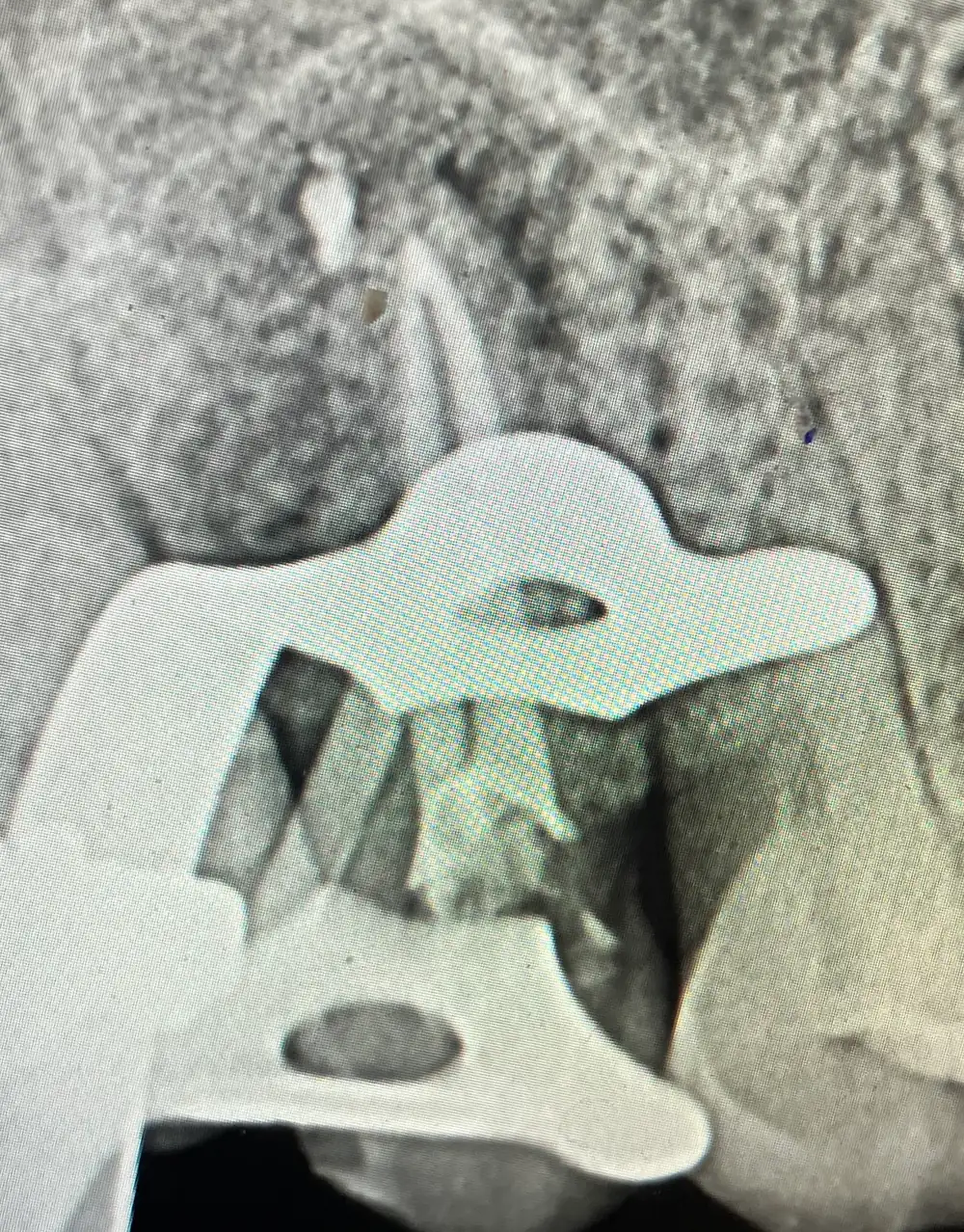
Isolated during treatment
Taken mid-treatment. The clamp holds a rubber dam that keeps the tooth dry and sealed off from saliva while the canals are cleaned.
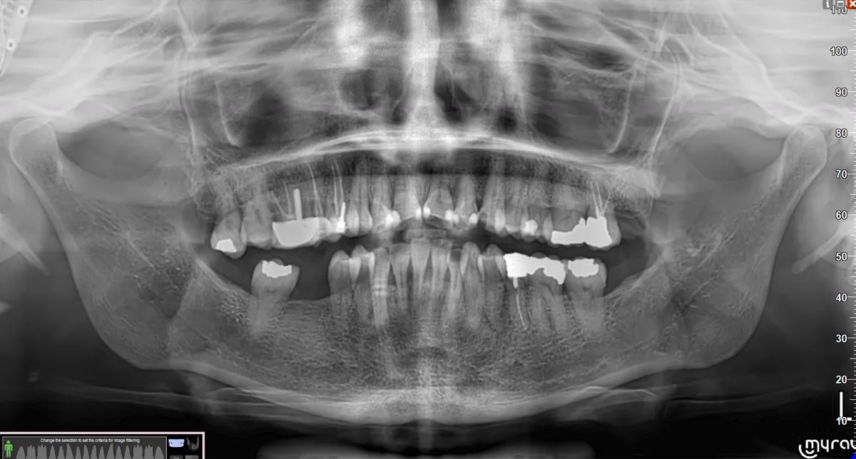
Full-mouth diagnosis
A panoramic X-ray taken at the clinic — how we find what's causing the pain before treating it.
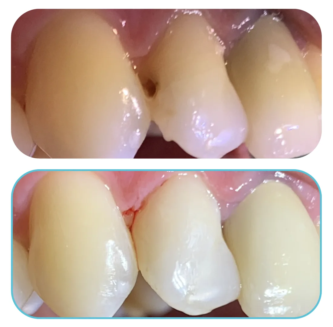
Decay on a front tooth
Decay between the front teeth, restored with a tooth-coloured composite filling.
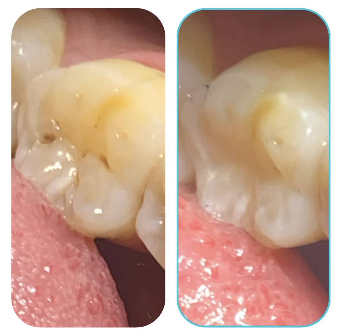
Decay in a molar
Decay on the biting surface of a back tooth, cleaned out and filled in one visit.
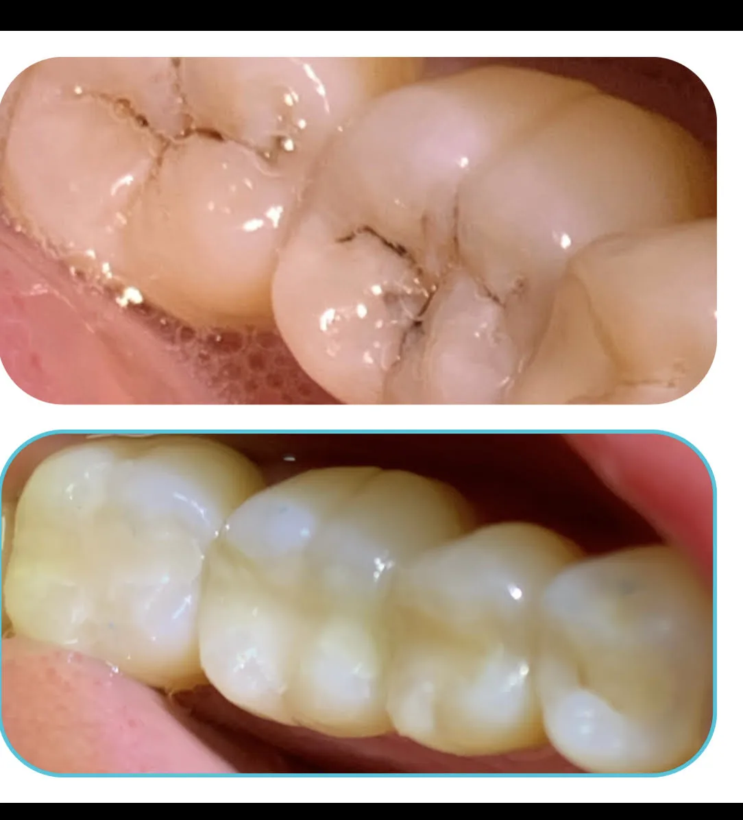
Stained molar grooves
Deep stained grooves on a molar, cleaned out and sealed before decay could spread.
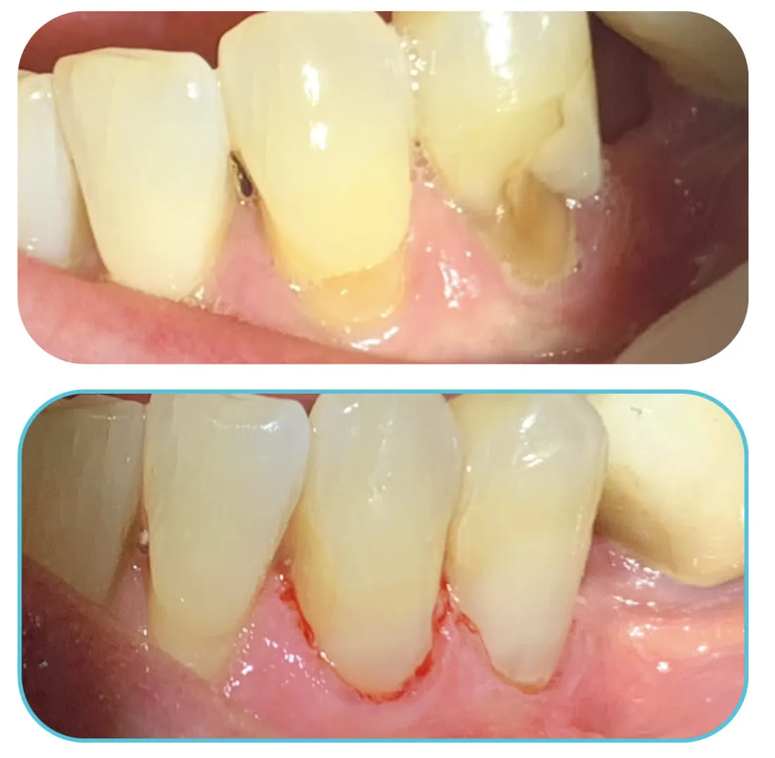
Wear at the gumline
Worn notches at the gumline — a common cause of cold sensitivity — restored with fillings.
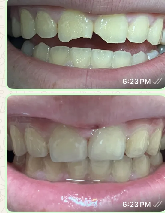
Chipped front tooth
A broken front tooth rebuilt in the clinic to match the shape and shade of the teeth next to it.
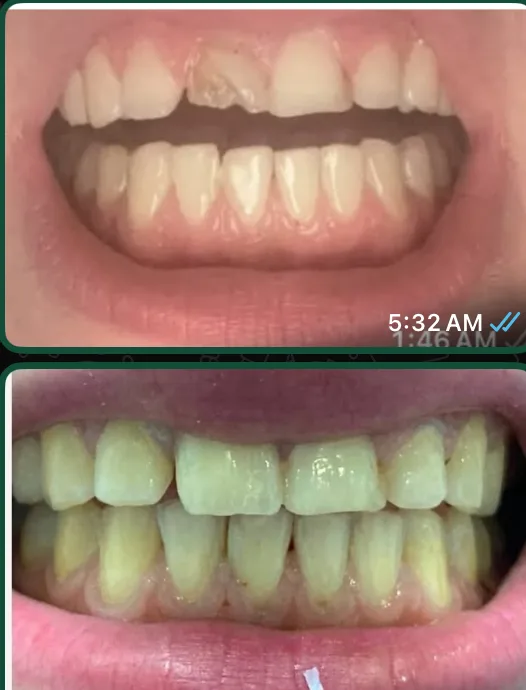
Worn front teeth
Chipped and worn upper front teeth, restored to an even edge.
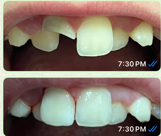
Rebuilding broken edges
Broken edges on the upper front teeth, rebuilt with tooth-coloured composite.
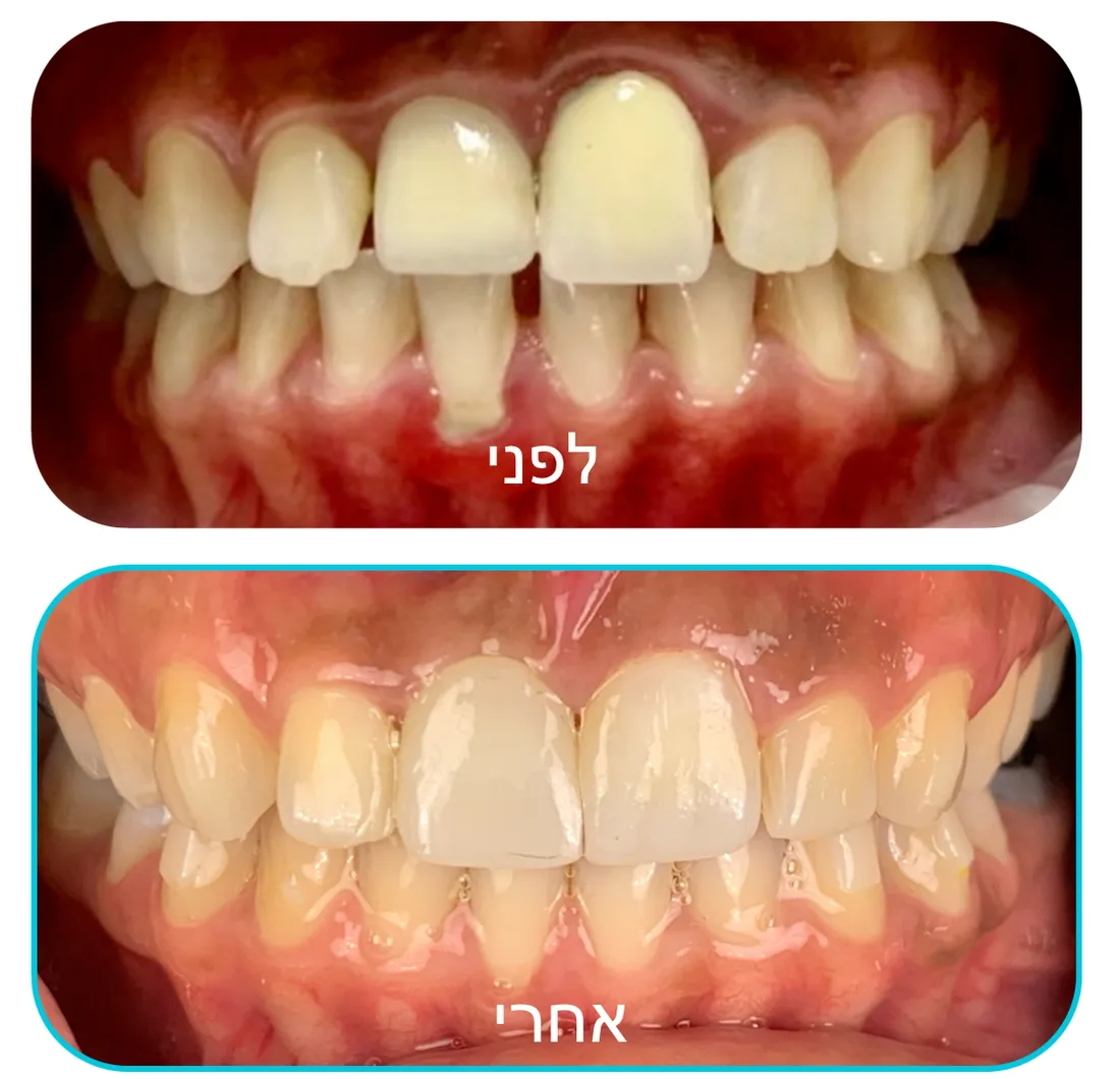
Crown on a front tooth
A damaged front tooth restored with a crown, shade-matched to the teeth beside it.
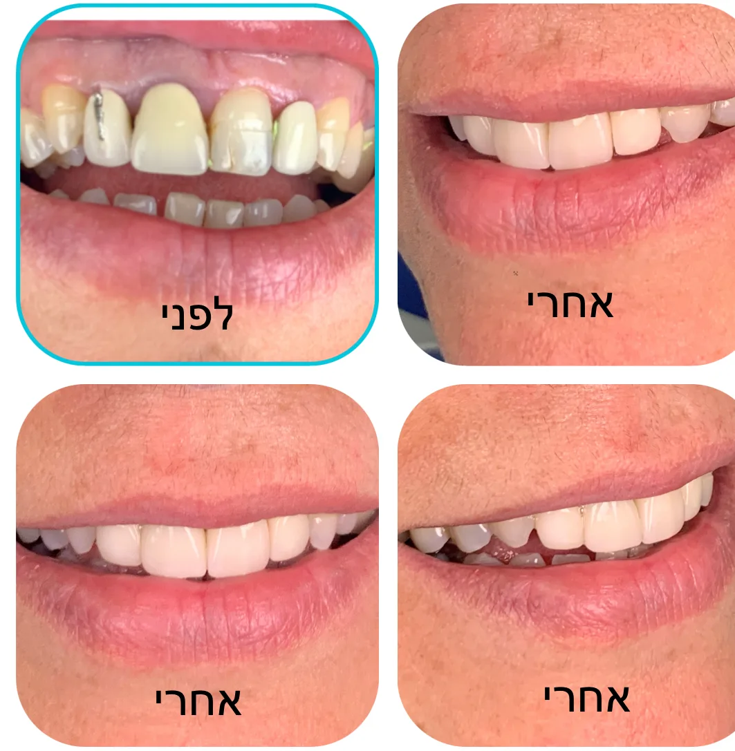
Rebuilding the front teeth
Broken and missing upper front teeth, rebuilt and shown from several angles.
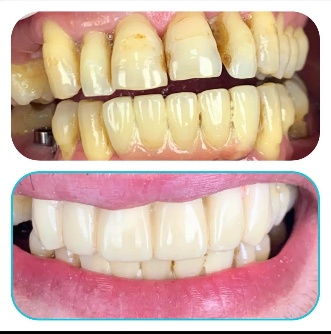
Heavily worn teeth
Years of wear and staining on the upper and lower teeth, restored across the arch.
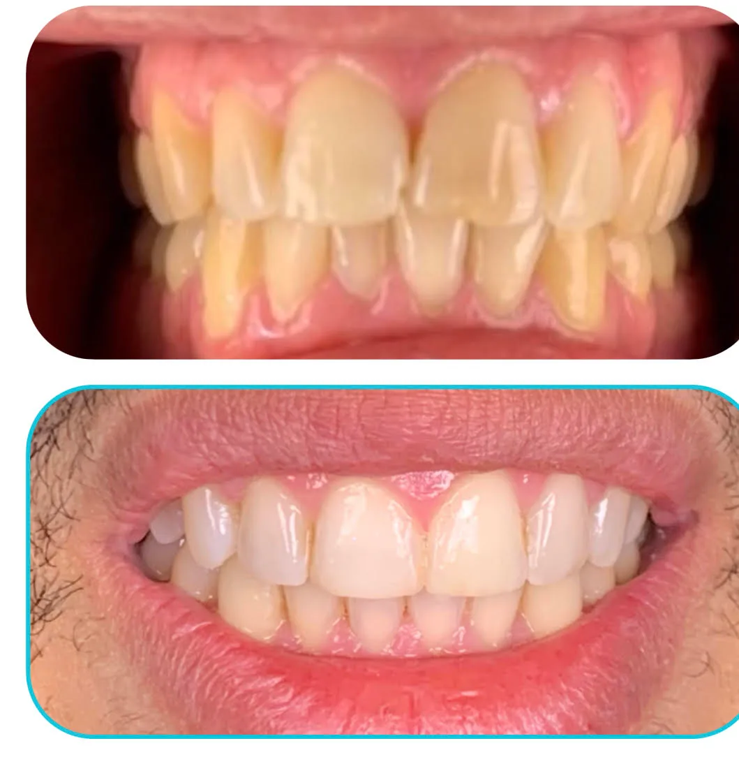
Upper and lower teeth
Worn and discoloured upper and lower teeth, restored.
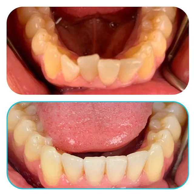
Lower front teeth
Build-up and staining on the crowded lower front teeth, cleaned and treated.
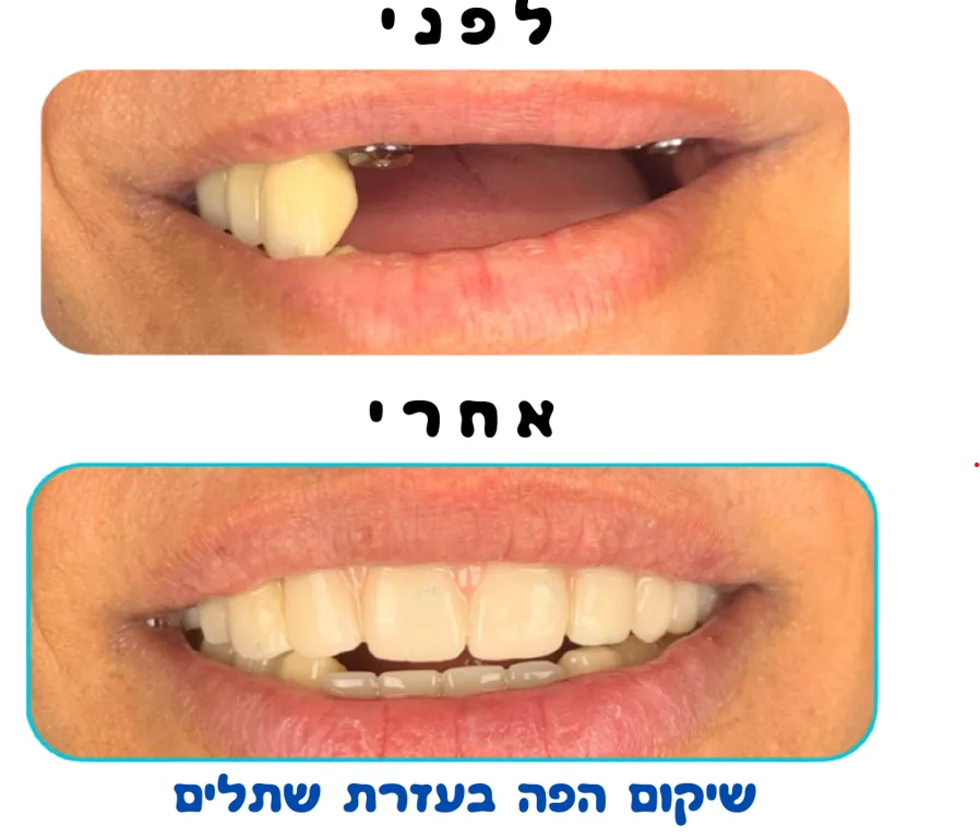
Rehabilitation with implants
Missing back teeth restored so the patient could chew on that side again.
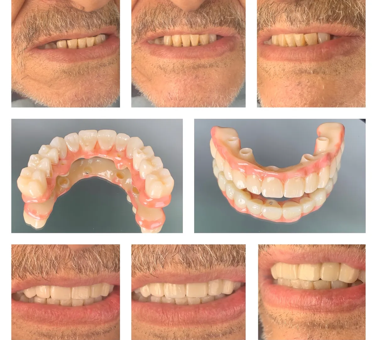
Full-mouth reconstruction
A full-mouth reconstruction, shown before, after, and with the finished work itself.
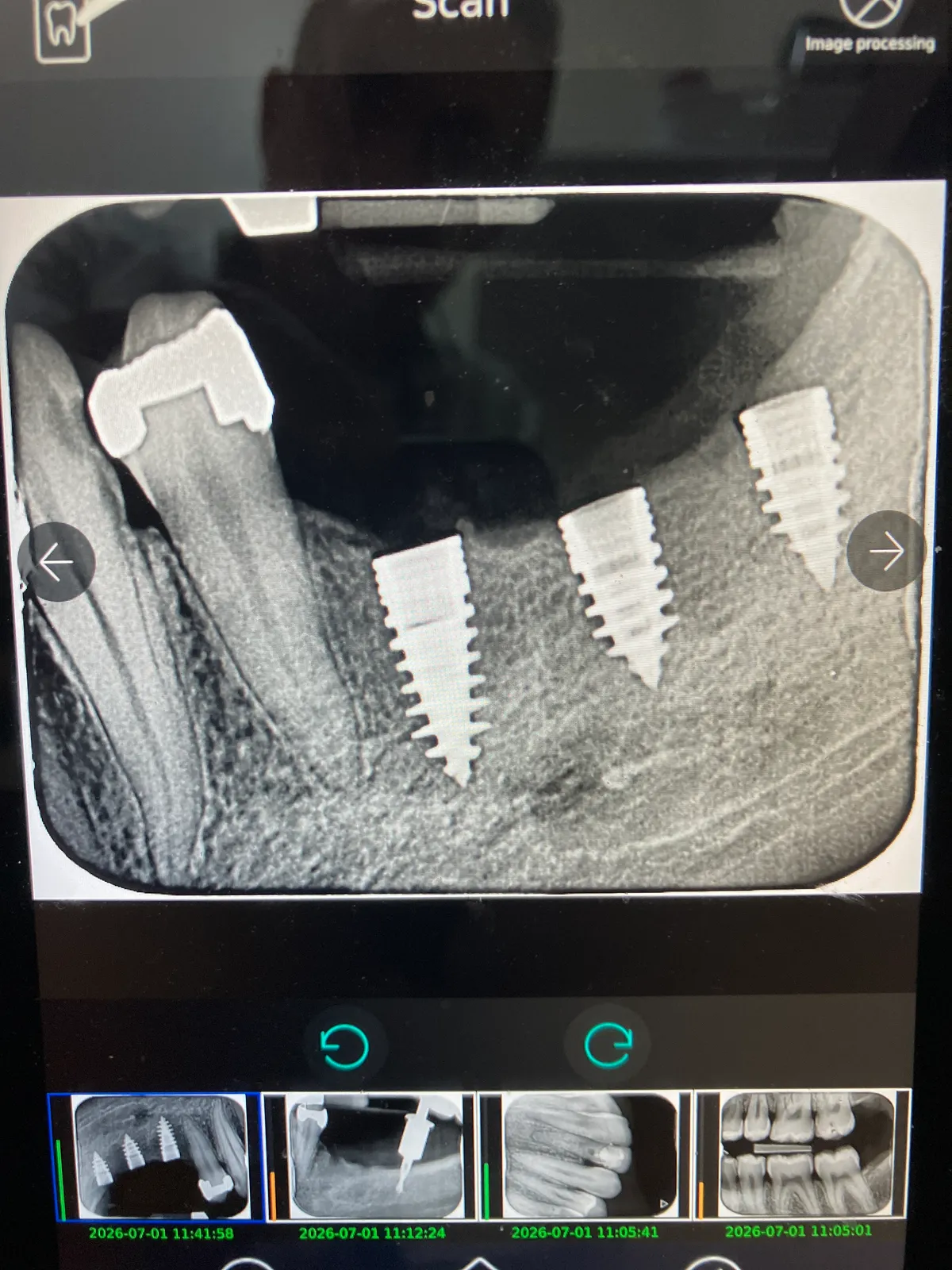
Implants on X-ray
Implants placed where teeth were missing, shown on the X-ray taken at the clinic.
Clinical images from our own patients, shared with permission. Every case is different — yours will be assessed on its own.
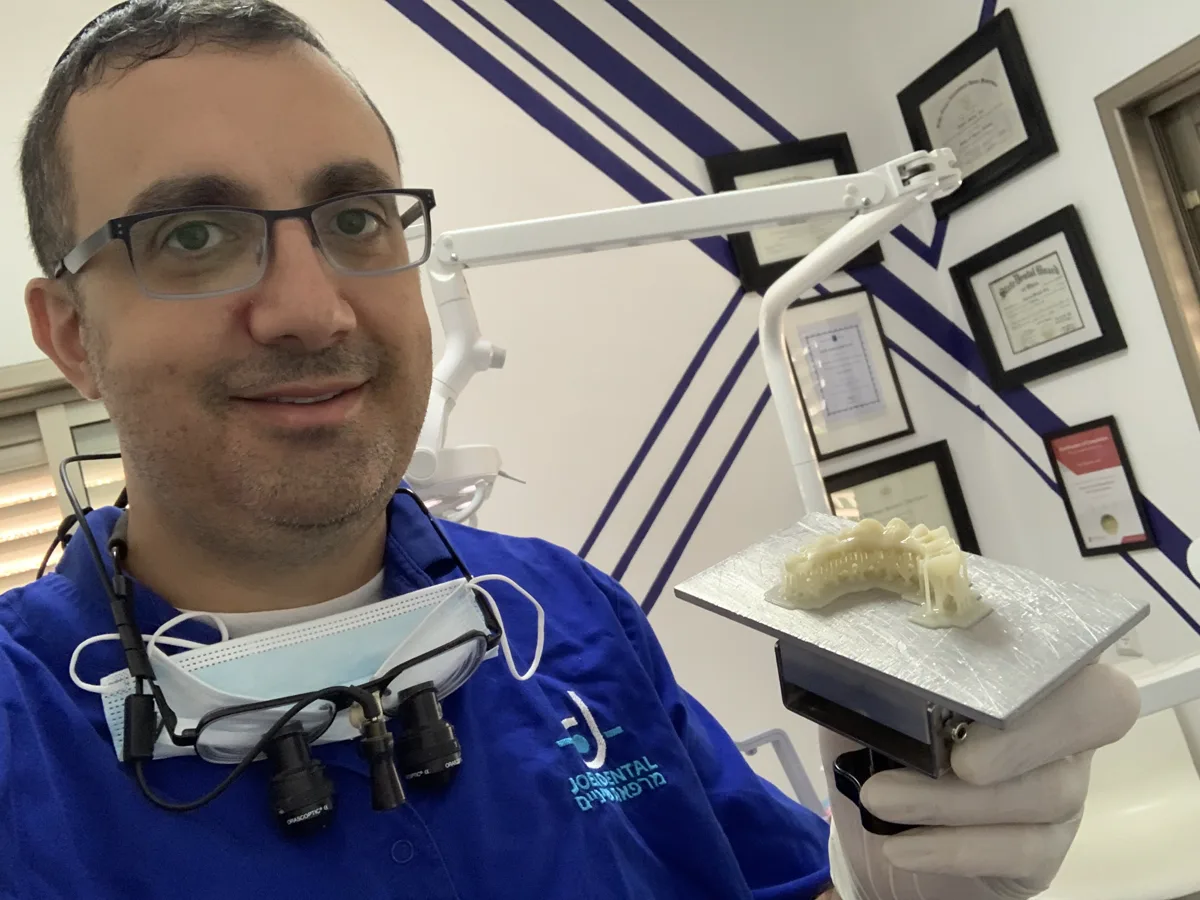
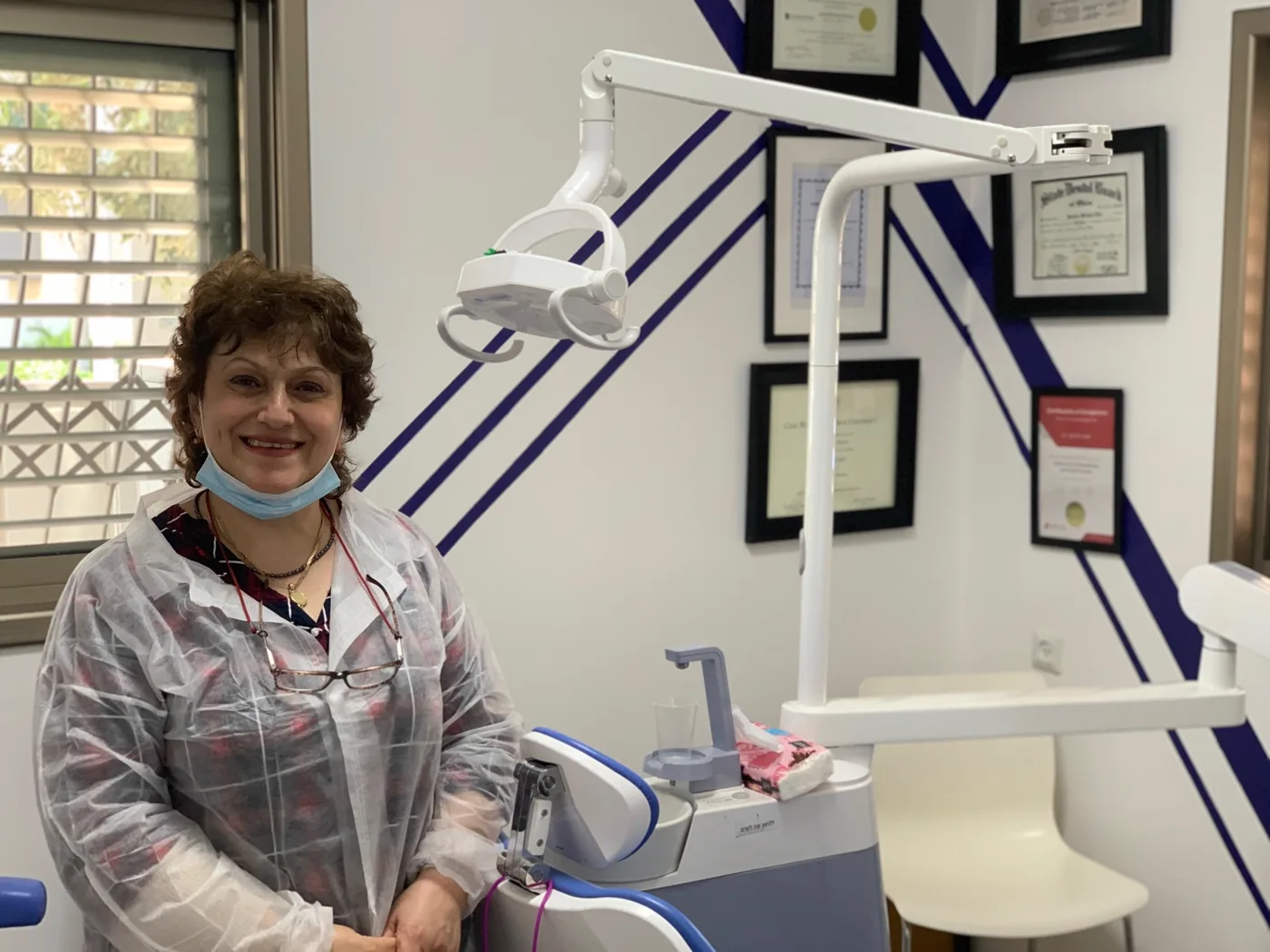
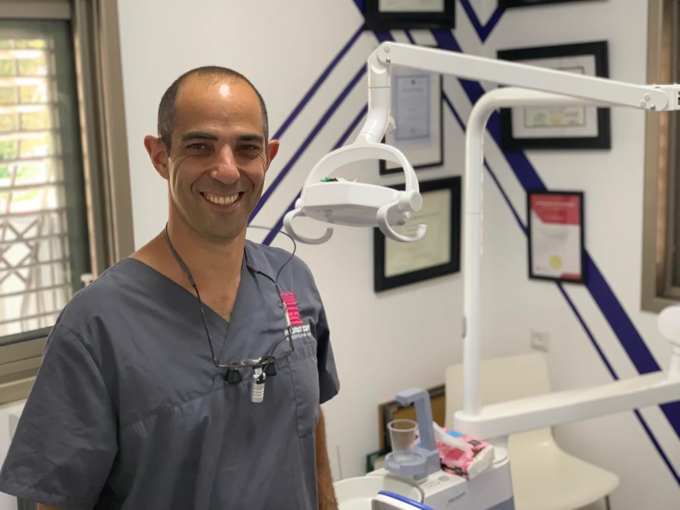
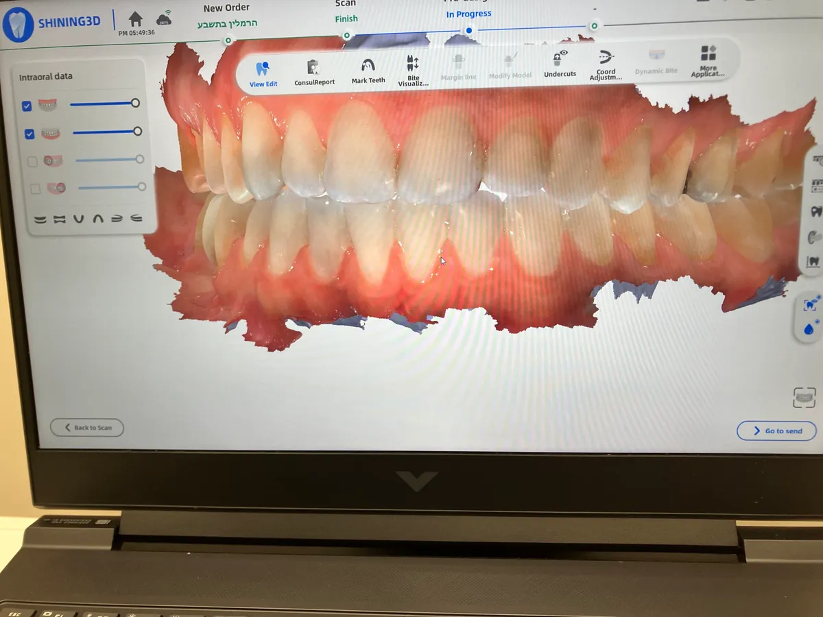
Inside the clinic
What patients tell their friends
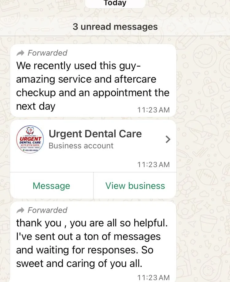
“The dentist that did my root canal on Wednesday at 1:45pm did a phenomenal job, and everyone was professional and kind.”
A recommendation passed between neighbours in Beit Shemesh.
WhatsApp NowTooth pain?
Let's fix it today.
Message Dr. Gabriel Joel, DMD directly on WhatsApp — no call centre, no sales script. We'll tell you what it'll cost before you sit in the chair.